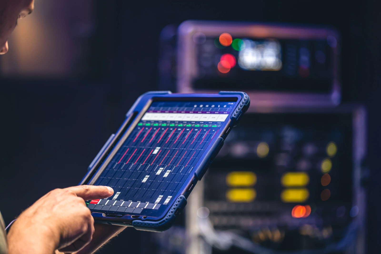
From 3 Minutes to 30 Seconds: How AI Rewired Plaque Detection for a Leading U.S. Cardiovascular Lab
Convert raw ultrasound scans into quantified arterial insights—automatically, accurately, and in under a minute.
No fatigue. No backlog. No bottlenecks.
Client
Leading U.S.-based cardiovascular diagnostics and research lab
Industry
Healthcare | Medical Imaging | Diagnostics
Timeline
6 weeks (from model development to deployment)
Scroll Down

Jump to Section
3 Min Read time
Quick Snapshot
A prominent cardiovascular lab faced a growing backlog of ultrasound images requiring manual review to detect plaque and measure arterial thickness. By implementing a deep learning model tailored for image segmentation and regression, the lab dramatically reduced diagnostic turnaround time, increased reviewer consistency, and enabled predictive risk scoring—without expanding its clinical team.
Brief
In cardiovascular imaging, manual reviews not only slowed operations but also introduced clinical inconsistency. Each scan took 2–3 minutes to analyze, and interpretations varied between radiologists. This affected throughput, increased cost per report, and left no room for predictive risk modeling.
Technostacks deployed an AI-powered solution that automated plaque segmentation and clinical metric extraction—delivering structured, predictive outputs in under 30 seconds per scan.
Challenges
Manual Processing Bottlenecks
Each scan required 180+ seconds of expert analysis, with increasing backlog and no scalable way to meet demand.
Inconsistent Accuracy
Inter-reader variability made plaque measurement and diagnosis inconsistent.
No Predictive Insights
The existing workflow offered no visibility into arterial aging or future cardiovascular risk.
High Per-Report Cost
Manual review models raised overhead, limited scalability, and affected operational margins.
Solution
An end-to-end AI pipeline was deployed to analyze ultrasound scans automatically and accurately.
Image Preprocessing
Normalized and standardized DICOM scans for optimal model input.
U-Net Segmentation
AI model identified arterial walls, plaque boundaries, and thickening zones.
Clinical Feature Extraction
Computed CIMT, plaque thickness, stiffness, and calcification metrics.
Regression-Based Risk Prediction
Estimated arterial age and progressive burden based on extracted features.
Web-Based Integration
Results integrated directly into the lab’s diagnostic interface for seamless review.
Technology used
- PyTorch + fastai for rapid deep learning model prototyping and optimization
- nnU-Net for automated, high-accuracy medical image segmentation
- OpenCV for image preprocessing, contour analysis, and feature extraction
- Albumentations for advanced data augmentation, improving model generalization
- Whisper STT for automated transcription and integration of clinician voice notes into diagnostic reports
Impact

Conclusion
This U.S.-based cardiovascular lab moved from manual interpretation to intelligent automation in just six weeks—delivering measurable clinical improvements without additional headcount. With faster reads, predictive insights, and consistent outputs, the lab redefined operational efficiency in diagnostics.
Ready to do the same?
Let’s discuss how AI can transform diagnostic imaging for your organization.
Our Solutions in Action
Read how we have transformed businesses along the way.
-
Enabling Smarter Industrial, Agricultural & Solar Operations through Long-Range, Low-Power IoT Monitoring with LoRa® Technology
From noisy factories to sprawling farmlands and solar fields, reliable real-time monitoring was once a distant dream. Traditional wired and Wi-Fi systems failed to cover vast areas or withst…
-
How a Jewellery Technology Pioneer Achieved 99% Accurate Diamond Testing with Technostacks AI-Powered Screening App
Clarity isn’t just for diamonds. With Technostacks AI-enabled mobile app, a jewellery technology innovator transformed diamond screening into a faster, more accurate, and fully automated e…

-
How a Chemical Manufacturing Giant Cut Costs by 60% and Accelerated ERP Deployment with Technostacks Zoho Implementation
Growth doesn’t have to wait. With Technostacks certified Zoho expertise, a global chemical leader turned years of ERP setbacks into measurable wins: faster deployment, lower costs, and AI-…
-
Reduce Order Errors. Cut Manual Work. Serve More Customers Without Lifting a Finger
Reduce human intervention in restaurant bookings by 90%, while giving every caller a smooth, intelligent ordering experience without hiring another soul.

-
Automated Answers, Zero Wait Time – Reducing HR Load with a RAG-Based Fleet Chatbot
Turn hours of repetitive HR and ops support into a single chatbot, one that knows every fleet policy, SOP, and HR guideline your team could ask for. It works 24/7, never gets tired, and unde…

-
Modernizing Balikbayan – Automating Last-Mile Delivery for Faster, Smarter Shipping
Sending a Balikbayan box from Sydney to Manila can be seamless, tracked from the moment it’s picked up at your doorstep to the second it reaches your loved one’s hands, all without the h…

-
Driving Performance, Resilience, and $1M Annual Savings Through Strategic Database Modernization
Improving system performance by 23%, cutting downtime incidents by 80%, and saving nearly $1 million annually, all while future-proofing critical infrastructure for a multi-business global e…

-
Executive hiring, reimagined: 90% faster CV reviews, zero compliance errors
Parsing resumes, aligning stakeholder input, and generating compliant, role-specific documents, all done faster than your next coffee break.

-
Digitizing Frost Protection – BLE-Powered, Mobile-First, Error-Proof
Configuring and managing heating cables across sub-zero zones without ever touching a control panel. That’s what we delivered: a fully connected, mobile-powered frost protection system tha…

Lets Talk
Have a challenge?Let us know.